Diagram Of Shoulder Joint - How Does The Shoulder Work Informedhealth Org / The shoulder joint is composed of three bones:
byAdmin•
0
Diagram Of Shoulder Joint - How Does The Shoulder Work Informedhealth Org / The shoulder joint is composed of three bones:. The rotator cuff muscles are important stabilizers and movers of the shoulder joint. It is the major joint connecting the upper limb to the trunk. The shoulder isn't just one bone, it's actually made up of three different bones and various tendons, ligaments, and muscles.the three bones located in the shoulder are the humerus, the scapula, and the clavicle. Dissection image of cartilage of glenohumeral joint in green. Capsule surrounds and attaches to humerus, outer circumference of glomerular fossa or labrum, scapula and head of biceps.
The partner should be noting the range and quality of motion of the shoulder joint. It is this joint that most people commonly think of as the shoulder joint. Diagram shoulder muscles 115 muscles of the pectoral girdle and upper limbs anatomy and. Motion usually occurs around joints. Other important bones in the shoulder include:
Shoulder Joint Illustrated And Annotated With Components On White Stock Vector Image Art Alamy from c8.alamy.com #tcml #anatomy #charsi #shoulderjoint #diahram #mbbslike, comment, share, subscribefor any query tell me in comment section. The acromioclavicular (ac) joint is located between the acromion (part of the scapula that forms the highest point of the shoulder) and the clavicle. A frozen shoulder diagnosis is made by observing the specific shoulder moving through a range of motion. The most flexible joint in the entire human body, our shoulder joint is formed by the union of the humerus, the scapula (or shoulder blade), and the clavicle (or collarbone). Glenohumeral joint (articulatio glenohumeralis) the glenohumeral, or shoulder, joint is a synovial joint that attaches the upper limb to the axial skeleton. Diagram muscle shoulder joint (page 1) 2. The shoulder isn't just one bone, it's actually made up of three different bones and various tendons, ligaments, and muscles.the three bones located in the shoulder are the humerus, the scapula, and the clavicle. Atlas of the anatomy of the joint of the shoulder on a ct arthrogram in axial, coronal, and sagittal sections, on a 3d images and on conventional athrogram.
It is one of the most mobile joints in the human body, at the cost of joint stability.
The rotator cuff muscles are important stabilizers and movers of the shoulder joint. Most relevant best selling latest uploads. There are three true joints in the shoulder girdle. The human shoulder is the most mobile joint in the body. Continued bursitis is when the bursa (a small sac filled with fluid that protects your rotator cuff) gets irritated. The shoulder joint is composed of three bones: Labeled anatomy chart of male triceps and back muscles on white background labeled human anatomy diagram of man's arm, shoulder and upper back muscles in a posterior view on a white. Acting in conjunction with the pectoral girdle, the shoulder joint allows for a wide range of motion at the upper limb. This is a tutorial on the glenohumeral joint.the glenohumeral joint is, as the name suggests a joint between the head of the humerus and the glenoid cavity of the scapula. There are a variety of muscles that have to work in concert to ensure the shoulder joint tracks properly with everyday activities. A frozen shoulder diagnosis is made by observing the specific shoulder moving through a range of motion. Diagram muscle shoulder joint (page 1) 2. This mobility provides the upper extremity with tremendous range of motion such as adduction, abduction, flexion, extension, internal rotation, external rotation, and 360° circumduction in the sagittal plane.
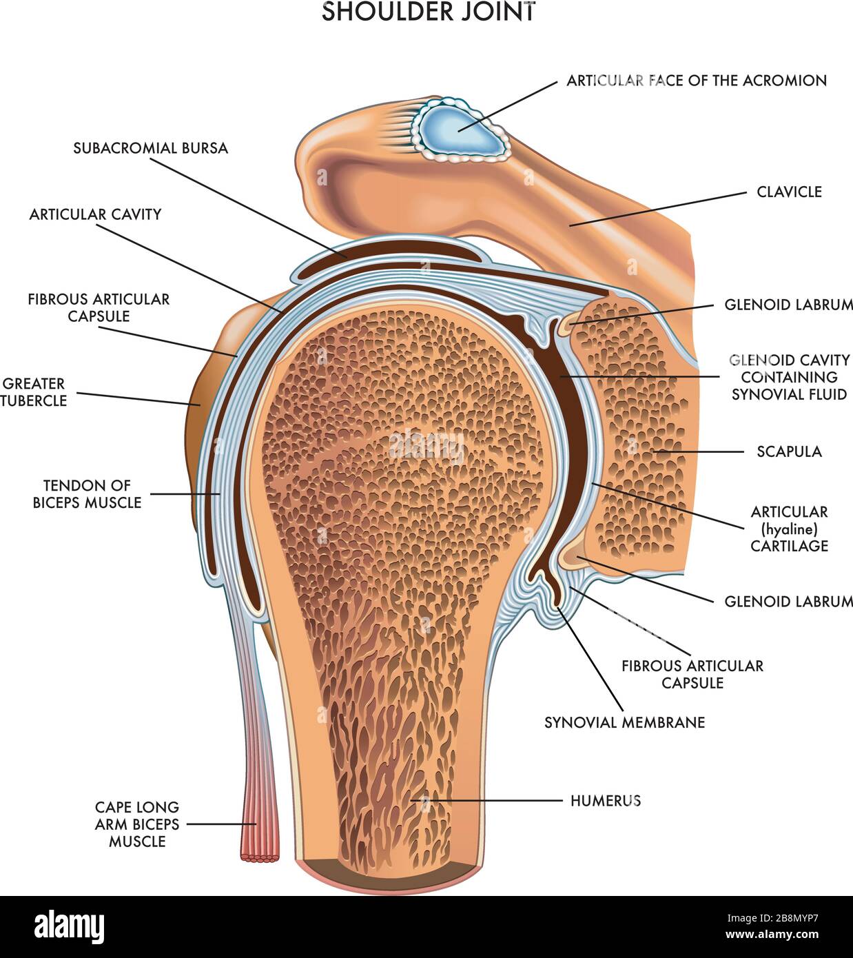
This page is about diagram muscle shoulder joint,contains upper extremity occupational therapy 205 with teresa at tufts university,2. It has to move through more than 180 degrees of motion in many directions, rotate, slide, and spin. Let's start by the anterior view of the diagram. License image this diagram illustrates the interior of the right shoulder joint capsule as viewed from the side. Dissection image of cartilage of glenohumeral joint in green.
What Makes The Shoulder Joint Different From Other Ball And Socket Joints How The Unique Structure Of The Shoulder Bursitis Shoulder Bursitis Shoulder Anatomy from i.pinimg.com This mobility provides the upper extremity with tremendous range of motion such as adduction, abduction, flexion, extension, internal rotation, external rotation, and 360° circumduction in the sagittal plane. Shoulder muscle and ligament diagram. A joint is formed where two or more bones meet. Continued bursitis is when the bursa (a small sac filled with fluid that protects your rotator cuff) gets irritated. Of these, the glenohumeral joint is the most important for shoulder motion. Two joints facilitate shoulder movement. Motion usually occurs around joints. The supraspinatus, infraspinatus, teres minor and subscapularis muscles are known as the shoulder rotator muscles.
The shoulder is the most complex joint in the human body.
Diagram shoulder muscles 115 muscles of the pectoral girdle and upper limbs anatomy and. See below to view an. This mobility provides the upper extremity with tremendous range of motion such as adduction, abduction, flexion, extension, internal rotation, external rotation, and 360° circumduction in the sagittal plane. Posted on january 20, 2015 by admin. You can see the glenoid cavity of the scapula here. Labeled anatomy chart of male triceps and back muscles on white background labeled human anatomy diagram of man's arm, shoulder and upper back muscles in a posterior view on a white. Capsule is made up of thin fibrous and areolar tissue. The most flexible joint in the entire human body, our shoulder joint is formed by the union of the humerus, the scapula (or shoulder blade), and the clavicle (or collarbone). Shoulder muscle and ligament diagram. It has to move through more than 180 degrees of motion in many directions, rotate, slide, and spin. License image this diagram illustrates the interior of the right shoulder joint capsule as viewed from the side. The shoulder joint (glenohumeral joint) is a ball and socket joint between the scapula and the humerus.it is the major joint connecting the upper limb to the trunk. Helps in internal rotation by allowing the individual to rotate the upper arm inwards and in.
Dissection image of cartilage of glenohumeral joint in green. A partner should observe you while moving the arm and shoulder. Two joints facilitate shoulder movement. Just like the muscle tissues in unique elements of the human physique, even our shoulder muscle tissues are prone to standard put on and tear. See below to view an.
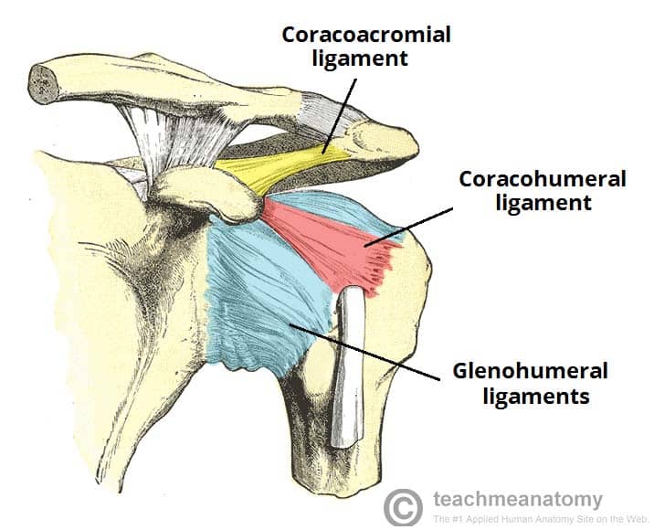
The Shoulder Joint Structure Movement Teachmeanatomy from teachmeanatomy.info Dissection image of coracohumeral ligament of glenohumeral joint in green. The shoulder isn't just one bone, it's actually made up of three different bones and various tendons, ligaments, and muscles.the three bones located in the shoulder are the humerus, the scapula, and the clavicle. Diagram muscle shoulder joint (page 1) 2. Or an acromioclavicular (ac) joint separation occurs when the joint of the clavicle and the scapula are disrupted. It is one of the most mobile joints in the human body, at the cost of joint stability. Diagram of the human shoulder joint, back view the left shoulder and acromioclavicular joints, and the proper ligaments of the scapula. A second joint in the shoulder is the junction of the collar bone with the shoulder blade, called the acromioclavicular joint. It includes a range of muscles such as front view of shoulder muscles.
The supraspinatus, infraspinatus, teres minor and subscapularis muscles are known as the shoulder rotator muscles.
It is one of the most mobile joints in the human body, at the cost of joint stability. Numerous muscles help stabilize the three joints of. #tcml #anatomy #charsi #shoulderjoint #diahram #mbbslike, comment, share, subscribefor any query tell me in comment section. Diagram shoulder muscles 115 muscles of the pectoral girdle and upper limbs anatomy and. Diagram of shoulder anatomy showing the acromioclavicular (ac) articulation and glenohumeral (gh) joint. Joint capsule is strengthened by multiple ligaments and tendons of shoulder joint. Just like the muscle tissues in unique elements of the human physique, even our shoulder muscle tissues are prone to standard put on and tear. It is this joint that most people commonly think of as the shoulder joint. Stand in front of a mirror. Of these, the glenohumeral joint is the most important for shoulder motion. A joint is formed where two or more bones meet. The bones of the pectoral girdle (clavicle and scapula) provide increased mobility to the. Beyond this, there is also a shoulder joint arrayed in a ball and socket formation, a rotator cuff, and various muscles like the deltoid muscle and the teres major muscle.
You can see the glenoid cavity of the scapula here diagram of shoulder. It is an extremely mobile joint, in which stability has been sacrificed for mobility.